
Trilobitology
Using 21st century visualization techniques on a -4.5*10^9th century trilobite.
EXPERIMENTAL BIOMECHANICS ON THE FRINGE OF C. TESSELATUS: WITH DR. KARLA HUBBARD AND DR. YOLANDA CRUZ
Oberlin College, Depts. of Geology and Biology
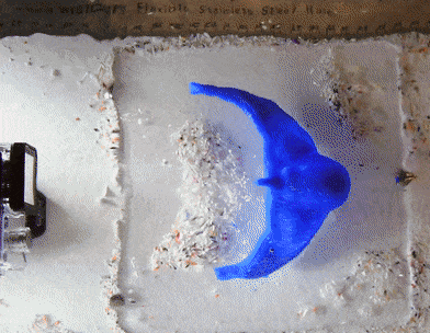
A 3D-printed CT scan of the trilobite C. tesselatus, showing the fringe's general inability to let fluid pass through.
tl;dr: About 500 million years ago, our oceans were crawling with a particularly weird animal--the trilobite Cryptolithus tesselatus. This creature is best identified by several dozen small holes in its head, a feature that paleontologists have been arguing about for over a century. In this study, we used several 3D printed trilobites to finally answer what those pits were for. We found that, contrary to popular belief, the holes were not used for filter feeding or physical strengthening. Instead, our results show that the unique pits were actually a growth signature, which allowed the trilobite's head to attain its especially large size.
The morphometric uniqueness of the trinucleid family of fossil arthropods, known as the trilobites, has led to a considerable amount of attention in paleontology literature. In particular, the distinctive hourglass-shaped pits that dot their anterior have been the subject of debate for over a century. Though anatomically well understood, their function remains unknown. Many proposals have been suggested, including its use as a sieve for filter-feeding, a strong shield for defense, and a sensory mechanism to compensate for their blindness. Despite the wide range of speculations, no study has attempted to model these hypotheses experimentally. Flume experiments and mechanical strength tests using a tenfold scale, 3D-printed model of a trinucleid head suggest that the dominant theories for over a century, filter-feeding and strengthening, are not well supported. It is proposed that the results suggest that the pits are an ontogenetic signature that optimize the cephalon’s growth to be maximal, providing trinucleids with an excellent mechanism for plowing through fine-grained silts and clays.
Non-linear ontogenetic shape change in Cryptolithus tesselatus (Trilobita) using 3-D geometric morphometrics: With Dr. Melanie Hopkins
American Museum of Natural History, Dept. of Paleontology
tl;dr: A common technique used by paleontologists is geometric morphometrics. If you take multiple snapshots of an animal as it grows, you can quantify how its shape changes by looking at how the shapes in the photographs change. The issue with 2-D morphometrics, however, is it loses all nuance in the third dimension. In this study, we used CT scans of a particular trilobite, Cryptolithus tesselatus, and noted how its shape changed during growth in three-dimensional space. From this nuance, we are able to make new inferences on the trilobite's paleoecology.
A decrease in the rate of cephalic shape change late in ontogeny has been documented for several species of trilobites, possibly associated with the cessation of segment release into the thorax. Qualitative descriptions of the ontogeny of Cryptolithus tesselatus Green, 1832, suggest that shape change in the cephalon was strongly influenced by the progressive accommodation of large funnel-shaped perforations (“fringe-pits”) over several molts. The number and arrangement of fringe-pits was established early in ontogeny, however, before thoracic segment release was completed. Due to the unusual and highly convex shape of the cephalon, we use three-dimensional (3D) geometric morphometrics to quantify shape change in this species and determine if there is a rate shift, and at what point in development this shift occurred. Three-dimensional morphometrics was made possible by extracting fixed and semi-landmarks from surface reconstructions of C. tesselatus rendered from CT scans of silicified specimens. Results show that the cephalon continued to change shape into adulthood, but that a threshold model with a rate shift associated with the cessation of new fringe-pits is best supported. 2D landmarks taken from the dorsal view fail to capture the dramatic change in convexity of the cephalon during development, but model comparison results are consistent with those based on the 3D landmark dataset, allowing comparison of this aspect of ontogenetic change with other species. Based on these comparisons, it appears that 1) trajectories are often better characterized by threshold models than simple linear regression models; 2) the timing of shifts may not be phylogenetically conserved.